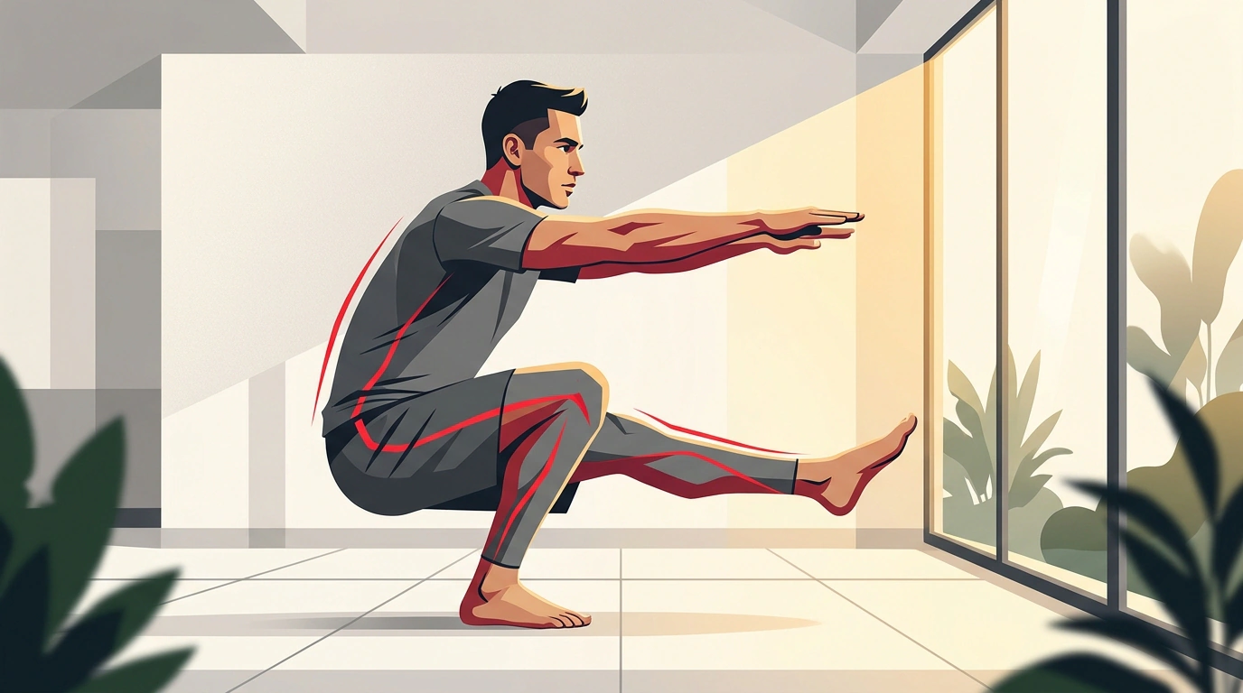
In gyms, the single-leg squat—the pistol squat—often carries an aura of sheer terror. Old-school trainers warn: "You'll wreck your knees, grind your menisci to dust, and shatter your kneecaps." And if someone gets an MRI out of curiosity and reads the radiologist's report, nine times out of ten they find a whole laundry list: "grade I–II osteoarthritis," "degenerative changes of the posterior horn of the medial meniscus," or a "Baker's cyst."
Seeing words like that, most people panic, quit exercising, buy elastic knee sleeves, and feel nervous even walking down stairs without holding the railing.
Yet modern sports orthopedics and evidence-based rehab live by a golden rule: we treat the person and their function, not the MRI image. Let's unpack why MRI findings aren't a death sentence for your joints, how deep-squat mechanics actually work, and how to safely build your legs up to demanding functional moves.
Scan vs. Function: Why 97% of Healthy People Have MRI Findings
Let's start with a surprising, rock-solid scientific fact. If you pull completely healthy adults over forty off the street—people who have never complained of knee pain—and put them in a high-field MRI scanner, what shows up?
A study in BMJ Open Sport & Exercise Medicine using a 3.0-Tesla MRI found radiological abnormalities in 97% of pain-free participants: cartilage thinning, horizontal meniscus tears, or bone marrow edema. A meta-analysis in the British Journal of Sports Medicine (BJSM) likewise showed signs of osteoarthritis on MRI in 19–43% of healthy people with zero joint pain.
Orthopedists call these "gray hairs and wrinkles on the inside." Degenerative changes are a normal marker of age and miles lived. A meniscus tear on a scan doesn't automatically mean your joint is falling apart.
The same goes for a Baker's cyst—a pocket of synovial fluid behind the knee. It's not a tumor and not a life sentence, just a secondary response by the joint capsule. As long as there's no acute flare-up, fluid circulates and clears naturally when your calf and thigh muscles pump regularly.
Joint Biomechanics: The Knee Caught Between Ankle and Hip
Anatomically, the knee is a simple hinge joint. It mostly bends and straightens, with only a tiny bit of rotation. Meanwhile, it sits trapped between two complex three-dimensional ball-and-socket-style movers: your ankle below and your hip above.
The knee is almost never the original culprit behind pain. It's usually the helpless victim of its neighbors' stiffness or weakness:
- Stiff ankle: If your Achilles tendon is tight and your ankle lacks bend (limited dorsiflexion), your center of mass shifts backward in a deep squat. To keep from falling over, your knee collapses inward (dynamic valgus), putting huge shear stress on the ligaments.
- Weak glutes: Your gluteus medius keeps your thigh from twisting inward. If it's weak, dropping down on one leg lets your femur rotate inward and pinches the meniscus between the bones.
Joint cartilage has no blood vessels of its own. In our piece on why loading protects your joints, we explained the "sponge" effect: cartilage only drinks in nutrients from synovial fluid when compressed and released during movement. Total rest starves the joint and speeds up degeneration. And in our look at painless knee clicking, we showed that popping sounds during squats usually come from harmless tiny gas bubbles collapsing.
From Regression to Progression: A Safe Path to Single-Leg Squats
So how do you work up to deep squats if you've never done them or worry about injury? Rehab specialists use regressions—ways to temporarily scale down the load:
- Foot and ankle mobility: rolling the sole on a firm ball, stretching your calves and soleus, and restoring ankle dorsiflexion.
- Glute activation: single-leg glute bridges and banded lateral hip abductions.
- TRX or gymnastic-ring squats: holding onto straps takes 40–50% of your body weight off the working leg, letting your brain and joint groove clean mechanics without fear or pain.
- Box squats: sitting back onto a sturdy bench or box and gradually lowering the target height.
- Eccentric lowers: lowering slowly on one leg for 3–4 seconds under control, then standing back up on both feet.
Ligaments and cartilage adapt much more slowly than muscles. Remodeling connective tissue takes months of steady, patient work without rushing.
Red Flags: When Training Through Pain Is Off-Limits
- Sharp, shooting, or stabbing pain deep inside the joint
- Sudden joint locking (the knee catches and you can't straighten your leg)
- Noticeable hot swelling with redness around the joint
- A feeling of instability (the knee gives way or buckles while walking)
When to Stop: Joint Red Flags
Joint adaptation doesn't mean blind heroics. Pain is your body's alarm system, and the type of pain matters.
Mild muscle soreness or gentle tendon tightness after a session is normal. But if you hit any symptom on the checklist above, stop training right away and see a sports orthopedist or orthopedic surgeon. You can also check our quick guide on why joints crack and where the real risks hide.
If there's no acute pain and your MRI just lists routine age-related wear, don't write off your knees. Smart movement, solid technique, and gradual progression restore joint strength and mobility better than any passive treatment.