Every day, thousands of people walk out of a cardiologist's office with the exact same look of bewilderment. On their blood chemistry panel, elevated cholesterol is highlighted in bold—say, low-density lipoprotein (LDL) at 3.8 or 4.2 mmol/L. The patient is around fifty, feels terrific, jogs in the mornings, has normal blood pressure, and nothing hurts. Then comes the verdict: "You need to take statins for life."
At that moment, a real storm erupts in a person's head. On one side is fear of cardiovascular catastrophes, heart attack, and stroke. On the other are forum myths about "wrecking the liver," muscle aches, and reluctance to start lifelong pills with zero symptoms. Even the doctor often hesitates: the patient sits in a borderline risk zone where formal calculators call for therapy, yet 100% certainty isn't there.
Cardiologist and science communicator Алексей Утин explains in detail how modern evidence-based cardiology resolves this agonizing dispute. Instead of reading statistical tea leaves, medicine today can peek inside the heart's coronary arteries in seconds using a simple low-dose CT scan and calculate the coronary artery calcium (CAC) score.
Let's unpack this elegant technology: how calcium forms in plaques, what the "Power of Zero" means, why statins paradoxically increase calcium density, and who truly needs this test.
Limits of Risk Calculators: Why SCORE2 Scales Misfire
In classic preventive cardiology, the decision to prescribe statins relies on predictive mathematical models. In Europe and Russia, that's the SCORE2 scale; in the US, the ASCVD Risk Estimator.
They work simply: the doctor enters your biological sex, age, smoking status, systolic blood pressure, and cholesterol level. The algorithm spits out a dry number: the probability of dying from a vascular event or having a heart attack over the next 10 years. If the risk exceeds 5–7.5%, clinical guidelines strongly advise starting lipid-lowering therapy.
Yet population calculators share one fundamental built-in flaw: they estimate abstract population statistics, while knowing nothing whatsoever about the personal anatomy of your own blood vessels.
In real clinical practice, this leads to two opposite errors:
- False overdiagnosis (overestimating risk in older adults). Picture a 62-year-old man. He has never smoked, keeps an ideal blood pressure of 115/75 mmHg, walks 10,000 steps a day, yet his LDL is slightly above average—3.4 mmol/L. By passport age alone, the SCORE2 scale automatically places him in the high-risk group. On paper, he qualifies for statins. Yet if we look inside his arteries, they may be completely clean, smooth, and free of atherosclerosis. The man spends years taking pills that change very little in his specific case.
- False underdiagnosis (underestimating risk in younger adults). The other extreme: a 42-year-old man with excess weight, a smoking history, and a family history (his father had a heart attack at 45). On the SCORE2 calculator, his risk comes out "green" and low—simply because he is still young. Yet at the ostium of his left coronary artery, a soft, unstable plaque may already be growing, ready to rupture during the next heavy strain at the gym.
Coronary calcium scoring changes the rules of the game completely. The scanner captures not theoretical probabilities, but the physical anatomical substrate of disease—already existing atherosclerotic vessel damage.
Biology of Vascular Calcium: How Soft Plaque Turns to Stone From Within
A massive number of everyday myths swirl around calcium in blood vessels. Some people fear drinking calcium-rich mineral water; others stop taking vitamin D for osteoporosis, seriously believing the mineral from their gut "crusts onto artery walls" like scale in a kettle.
The physiology of atherosclerosis works completely differently. Coronary artery calcification is not a random salt deposit—it is a highly organized, biologically regulated wound-healing process. And it happens strictly inside a pre-existing atherosclerotic plaque.
Let's trace this chain step by step:
- Lipid infiltration into the intima. When the delicate inner lining of an artery (the endothelium) is injured by high blood pressure, cigarette smoke components, or homocysteine, atherogenic LDL and ApoB particles slip into the subendothelial layer—the arterial intima.
- Inflammation and foam cells. Inside the vessel wall, lipids oxidize. The immune system treats them as foreign invaders and dispatches macrophages. Macrophages greedily engulf oxidized cholesterol, become engorged, and turn into so-called "foam cells." When these cells die, a liquid, inflamed, necrotic lipid core forms in the vessel wall.
- Osteogenic cell transformation. To wall off the dangerous inflammatory focus from the arterial lumen, the body tries to entomb it. Under inflammatory signals (bone morphogenetic proteins BMP-2 and interleukins), vascular smooth muscle cells pull off a phenotypic trick: they transform into bone-like cells—osteoblasts.
- Hydroxyapatite crystallization. These cells begin secreting matrix vesicles that crystallize calcium phosphate—hydroxyapatite Ca10(PO4)6(OH)2—in the exact same way skeletal bone forms. First, microscopic specks of calcium appear in the plaque cap, gradually fusing into solid stony plates.
From this follows a fundamental law of vascular biology: calcium never exists in a healthy arterial wall. If a CT scanner detects calcium in coronary vessels, it proves unequivocally that an atherosclerotic plaque has already formed and developed at that spot.
Scanning Technology: 5 Seconds Without Contrast and the Agatston Scale
Many patients confuse a non-contrast coronary calcium CT with a complex CT coronary angiogram. The difference between them is fundamental:
- A calcium scan uses no intravenous contrast. No iodine-based dyes are injected into a vein, meaning zero toxic load on the kidneys and zero risk of allergic reactions.
- Scanning uses ECG gating. The X-ray tube captures images strictly during diastole—the brief moment when the heart muscle is completely relaxed and still. That eliminates motion blur.
- Minimal radiation exposure. The radiation dose is under 1 millisievert (mSv)—typically around 0.5–0.8 mSv on modern scanners. That is comparable to a standard chest X-ray or the natural background radiation each of us absorbs from the sun and space over a couple of months.
- The scan itself takes literally 3–8 seconds on a single short breath-hold.
How is the score calculated? In 1990, American cardiologist Arthur Agatston developed the mathematical algorithm that became the global gold standard—the Agatston CAC Score.
The CT scanner takes cross-sectional slices of the heart 2.5–3 millimeters thick. Software searches for foci along the four main coronary arteries: the left main, left anterior descending, circumflex, and right coronary artery. A calcified lesion is defined as an area with density above 130 Hounsfield units (HU) and an area of at least 1 mm². Each lesion's area is multiplied by a weighting factor for its peak density (from 1 to 4), and all points are summed into a single number.
Interpreting Results: From the "Power of Zero" to a Critical 400
With an Agatston score printout in hand, cardiologists rely on consensus clinical guidelines from the American Heart Association (AHA) and American College of Cardiology (ACC):
| CAC Score (points) | Degree of Calcification | Cardiovascular Risk | Clinical Strategy |
|---|---|---|---|
| 0 | No calcium ("Power of Zero") | Extremely low (< 1% events over 10 years) | Statins not needed; lifestyle; repeat in 3–5 years |
| 1 – 99 | Minimal calcification | Moderate risk | Individualized decision (statins if risk enhancers present) |
| 100 – 399 | Pronounced calcification | High risk | Mandatory statin initiation; target LDL < 1.8 |
| ≥ 400 | Severe arterial disease | Very high risk (CAD equivalent) | Maximum-dose statins + ezetimibe; stress testing |
The "Power of Zero" Phenomenon
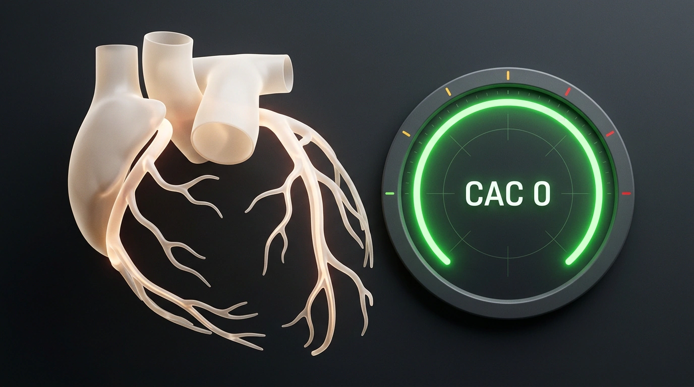
The most reassuring test result is CAC = 0. In evidence-based cardiology, this is called "the Power of Zero."
The landmark multi-year MESA (Multi-Ethnic Study of Atherosclerosis) followed more than 6,800 people for over a decade. The data showed that if a person has a calcium score of zero, their 10-year risk of dying from a heart attack is less than 1%, even if their cholesterol or blood pressure is moderately elevated.
Zero calcium gives doctor and patient a so-called "warranty period" of 3–5 years. That means starting statin medication can be safely deferred. The patient gains time and strong motivation: normalize weight, improve diet, quit smoking, and add physical activity without fearing a sudden vascular catastrophe.
If the score climbs past 100 points, doubts vanish: atherosclerosis is actively underway, vessels are already damaged, and statins are needed immediately to save lives. We have already detailed how new clinical guidelines for fighting high cholesterol work, and the calcium score has become their key referee.
The Calcification Paradox: Why Statins Increase Plaque Density
Now brace yourself for the main intellectual paradox that regularly trips up not only patients, but also inexperienced doctors.
3D model of a coronary artery cross-section with a stable mineralized plaque and unobstructed blood flow
Imagine this scenario: a cardiologist prescribes a modern statin (atorvastatin or rosuvastatin) to someone with a calcium score of 150. The patient takes the pills faithfully for a year, retests their lipid panel—LDL cholesterol drops beautifully from 4.2 to a target 1.6 mmol/L. Pleased, the patient goes for a repeat CT scan to confirm the "plaques melted away"... and is horrified to see their coronary calcium score rose from 150 to 190!
The first thought: "The pills aren't working—they only made things worse!" In reality, the exact opposite happened: the medication worked to perfection. In science, this phenomenon is called The Calcification Paradox.
Why do statins raise coronary calcium?
- The main danger of atherosclerosis isn't plaque size itself, but plaque instability. A heart attack doesn't happen because a hard plaque slowly narrows the lumen; it happens when a soft, inflamed plaque with a thin fibrous cap suddenly ruptures. Platelets instantly pile onto the tear, a clot forms, and the artery slams shut in seconds.
- Statins have potent anti-inflammatory effects. They pull liquid cholesterol out of the plaque, suppress tissue-degrading enzymes, and stimulate dense mineralization of the plaque with calcium.
- Statins turn a soft powder keg into a monolithic, stable stone scar. A calcified plaque can no longer rupture!
- On statin therapy, the calcium score naturally rises by 10–25% per year due to densification of existing plaques.
From this flows an ironclad clinical rule: never do a repeat calcium CT to check whether statins are working. Once medication has started, the test loses all diagnostic meaning. Statin effectiveness is tracked strictly through blood tests for LDL and ApoB, as we noted in our review of evidence-based medications against cardiovascular risk.
Who It Saves and Who Doesn't Need It: Boundaries of the Test
Like any high-precision medical tool, a coronary calcium CT has strict boundaries of usefulness. This test was built for a specific clinical question and is completely useless when ordered inappropriately.
Who Is the Ideal Candidate?
Asymptomatic men and women aged 40–75 in the intermediate or borderline risk category (5–10% on SCORE2):
- when cholesterol is elevated, but other obvious risk factors are few;
- when a patient strongly hesitates to start statins and wants objective proof;
- when there is a family history of early heart attacks in parents, yet formal personal risk still looks low.
Who Definitely Does NOT Need the Test?
- Patients with an established diagnosis of cardiovascular disease. Anyone who has already had a heart attack, stroke, stenting, or bypass surgery, or has carotid artery stenosis over 50%, is ALREADY in the extreme-risk group. Statins are vitally indicated regardless of any CT number. Spending time and taking radiation makes no sense.
- Patients with type 2 diabetes over age 40 or severe familial hypercholesterolemia (LDL above 4.9 mmol/L). In these groups, baseline risk is already high, and therapy is started without hesitation.
- Patients with angina symptoms (chest pain on exertion, shortness of breath). A calcium CT is strictly for people WITHOUT symptoms. If pain is already present, contrast angiography or stress echocardiography is needed.
- Young smokers under age 40. At younger ages, plaques may be purely lipid-rich and not yet mineralized. The score can show a false zero despite an active process.
Coronary calcium scoring is a brilliant example of 21st-century personalized medicine. It spares unnecessary medication for those whose vessels are clean, and opens the eyes of those who underestimated hidden atherosclerosis in time.
If you are already taking statins prescribed by a cardiologist, don't waste money on a repeat coronary calcium CT: an increase in score during treatment is normal and reflects plaque stabilization, not worsening disease. Monitor therapy success by checking your LDL level at the lab.